Primary mouse microglia cells are essential in studying the central nervous system (CNS), particularly in understanding neuroinflammation, neurodegenerative diseases, and brain homeostasis. Microglia are the resident immune cells of the CNS, responsible for maintaining brain health by responding to injury, clearing pathogens, and regulating neuronal connections. Research on primary mouse microglia has been pivotal for deciphering the cellular and molecular mechanisms underlying various neurological conditions.
Characteristics of Microglia Cells
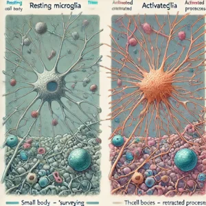
Microglia are characterized by their small cell bodies and extensive, highly motile processes, which constantly monitor the CNS environment. They originate from progenitor cells in the yolk sac during early embryonic development and migrate to the brain, where they populate throughout life. These cells play dual roles, both protective and destructive, depending on the context. Under normal conditions, microglia maintain synaptic plasticity and prune synaptic connections, but in response to injury or disease, they become activated, adopting a pro-inflammatory or anti-inflammatory phenotype. Understanding these dynamic changes has been facilitated by studies using primary mouse microglia derived from brain tissue.
Isolation and Culturing Techniques
Isolation of primary mouse microglia involves harvesting brain tissue, enzymatic digestion, and mechanical dissociation to obtain a single-cell suspension. The cells are then purified using techniques such as immunopanning or magnetic-activated cell sorting (MACS). Cultured primary microglia provide a robust model for in vitro studies, allowing researchers to examine microglial behavior, signaling pathways, and responses to various stimuli. For protocols and best practices, you can refer to resources provided by academic institutions such as Stanford University, University of California, San Francisco, and National Institutes of Health.
Research Applications
- Neurodegenerative Diseases: Primary mouse microglia are widely used to study diseases like Alzheimer’s, Parkinson’s, and multiple sclerosis. These cells help elucidate the role of neuroinflammation and the immune response in disease progression. More information on this can be found on NIA Alzheimer’s Disease Resource Center.
- Brain Development and Injury: Microglia play a crucial role in brain development, synaptic pruning, and recovery from injury. Research funded by NIH and studies conducted at Harvard University have been instrumental in revealing these functions.
- Immunological Responses: As the brain’s primary immune cells, microglia are essential for understanding CNS infections and immune responses. Studies available from CDC and Johns Hopkins University have focused on the role of microglia in response to viral infections.

Microglial Activation States
Microglia can switch between various states, primarily:
- Resting (Surveying): In a non-activated state, microglia constantly surveil the brain environment, clearing debris and supporting neuronal health.
- Activated (Reactive): Upon encountering pathogens or damage signals, microglia become activated, releasing cytokines, chemokines, and other signaling molecules. Activation can be either pro-inflammatory (M1) or anti-inflammatory (M2), a classification that has guided much research, particularly in studying neurodegenerative diseases. Studies from NIH and Yale University have shown how these states affect brain function.
Recent Advances in Microglia Research
Recent advances include the development of transgenic mouse models, enabling more precise manipulation and tracking of microglia in vivo. These models help study microglial responses in a living organism, providing insights into disease mechanisms that cannot be captured through cell cultures alone. Research institutes such as Massachusetts Institute of Technology (MIT) and University of California, San Diego (UCSD) are at the forefront of developing these technologies.
Cultured Primary Mouse Microglia vs. Cell Lines
While cell lines like BV2 microglia are widely used due to their convenience, they do not fully replicate the characteristics of primary microglia. Primary microglia maintain the phenotype and responses seen in vivo, making them a more accurate model for research. Studies comparing these models are available on PubMed and reports from University of California, Berkeley.
Primary mouse microglia cells serve as a critical tool for understanding the CNS’s complex network, especially the immune responses, disease mechanisms, and therapeutic potentials in neurodegenerative conditions. Ongoing research continues to reveal the multifaceted roles of microglia, contributing to the development of new therapeutic strategies targeting brain diseases. For further reading, refer to the National Library of Medicine, Centers for Disease Control and Prevention, and National Institute on Aging.
By leveraging resources from educational and government institutions, researchers can continue to expand our understanding of these vital CNS cells, ultimately leading to breakthroughs in treating various neurological disorders.