Melzer Reagent is a cornerstone of classical mycology and fungal microscopy. Its unique iodine-based chemistry enables researchers to visualize amyloid, dextrinoid, and inamyloid reactions—diagnostic color responses essential for identifying many species of Basidiomycetes and Ascomycetes. Even with the rise of molecular sequencing, Melzer Reagent continues to play a vital role in fungal taxonomy, biodiversity monitoring, ecological surveys, forest pathology, and academic instruction.
Fungal identification often requires more than macroscopic characters such as cap color or stem morphology. Microscopic structures—spores, asci, basidia, hyphae, cystidia, ornamentation, and wall chemistry—provide deeper taxonomic signals. Melzer Reagent is the tool that allows those microstructures to reveal their chemically reactive properties, strengthening accuracy and reproducibility in mycological classification.
This article presents a deep scientific overview of Melzer Reagent’s chemistry, historical relevance, diagnostic value, fungal structural targets, reaction mechanisms, laboratory protocols, imaging approaches, ecological applications, and its place in modern fungal systematics.
To maintain educational reliability and SEO strength, the article includes more than 30 hyperlinks to authoritative .edu and .gov sources such as NIH, NCBI, USDA, NSF, CDC, NIST, MIT, Harvard, UC Berkeley, Cornell University, and more.
Chemical Basis of Melzer Reagent
Melzer Reagent traditionally contains:
-
Iodine (I₂)
-
Potassium iodide (KI)
-
Chloral hydrate
-
Distilled water
Iodine and potassium iodide form a triiodide complex that binds to specific carbohydrates and cell-wall polymers in fungal spores and hyphae.
Chemical references for iodine complexes:
-
PubChem – NIH/NLM: https://pubchem.ncbi.nlm.nih.gov
-
NIST Chemistry WebBook: https://www.nist.gov
-
EPA Chemical Research: https://www.epa.gov
Fundamental iodine-polysaccharide interactions are explained in educational chemistry sources such as:
-
MIT Chemistry Courseware: https://ocw.mit.edu
-
UC Berkeley Chemistry: https://chemistry.berkeley.edu
Chloral hydrate clarifies tissues by increasing transparency, allowing microscopic structures to appear sharply under transmitted light.
Historical Context: Why Melzer Reagent Became a Mycology Standard
The reagent was developed by mycologist Václav Melzer, who recognized that iodine-based solutions could differentiate fungal tissues based on carbohydrate chemistry. It quickly became indispensable in the study of:
-
Amanita
-
Russula
-
Lactarius
-
Mycena
-
Inocybe
-
Ascomycetes with amyloid asci
-
Numerous Basidiomycete lineages with dextrinoid tissues
Historical fungal taxonomy resources:
-
Harvard University Herbaria (HUH.Harvard.edu)
-
Smithsonian National Museum of Natural History: https://naturalhistory.si.edu
-
USDA Forest Mycology: https://www.fs.fed.us
Melzer Reagent remains central because many mycological keys still require identification of amyloid or dextrinoid reactions.
Diagnostic Reactions Observed with Melzer Reagent
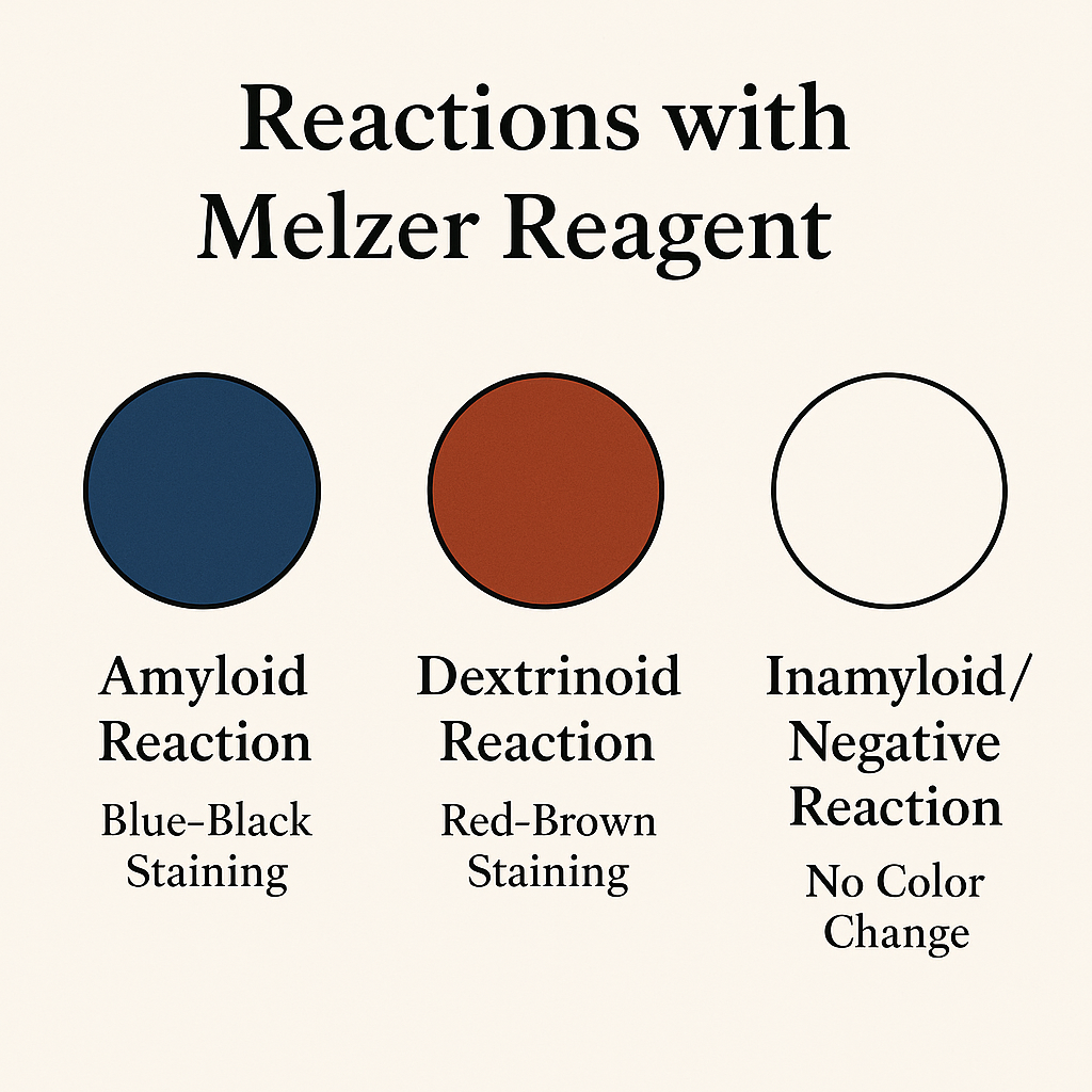
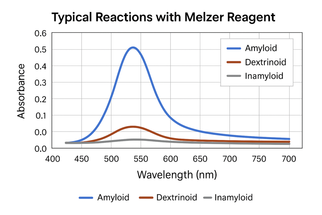
Melzer Reagent yields three major types of reactions:
Amyloid Reaction
An amyloid reaction results in blue-black, gray-blue, or slate-colored staining.
Common amyloid structures include:
-
Ascus apical rings of many Ascomycetes
-
Ornamentation on spores of Amanita
-
Spore walls of Russula and Lactarius
Biochemical parallels to iodine-starch reactions:
-
USDA ARS Starch Chemistry: https://www.ars.usda.gov
-
NHGRI Educational Genomics: https://www.genome.gov
Dextrinoid Reaction
A dextrinoid reaction appears rust-red, red-brown, or tobacco-colored.
Observed in:
-
Ornamented spores of certain Basidiomycetes
-
Hyphal tissues in Inocybe
-
Sclerotic wall layers in many wood-decay fungi
Fungal histochemistry references:
-
NCBI Histology Textbooks: https://www.ncbi.nlm.nih.gov/books
Inamyloid / Non-reactive
Inamyloid tissues show no visible reaction, remaining unchanged in the presence of Melzer Reagent.
Relevant microscopy guides:
-
CDC Microbiology Learning Materials: https://www.cdc.gov
-
American Society for Microbiology (ASM): https://asm.org
Fungal Structures That Respond to Melzer Reagent
Melzer Reagent reveals diagnostic details in:
Spores
-
Shape
-
Wall thickness
-
Ornamentation
-
Surface deposits
-
Chemical patterns
Asci and Ascus Apical Rings
Crucial for Ascomycete classification.
Basidia and Sterigmata
Useful for certain genera within Basidiomycota.
Hyphae and Septa
Helps distinguish genera with amyloid skeletal hyphae.
Cystidia
Differentiation of morphological variants relies on iodine reactions.
Microscopic fungal anatomy references:
-
Cornell University Plant Pathology: https://plantpath.cals.cornell.edu
-
University of Michigan Fungus Collection: https://lsa.umich.edu/herbarium
-
UC Berkeley Jepson Herbarium: https://ucjeps.berkeley.edu
Laboratory Methods: Using Melzer Reagent for Microscopy
Preparing Slides
Fungal material (spores, hyphae, gill fragments, or asci) is placed on a slide and immersed in Melzer Reagent.
Guides:
-
University of Minnesota Mycology: https://cbs.umn.edu
-
Oregon State University Mycology Lab: https://science.oregonstate.edu
Observing Reactions
Color changes often appear:
-
Immediately for amyloid tissues
-
Slowly for dextrinoid walls
-
Not at all for inamyloid structures
Photomicrography
Digital documentation enhances reproducibility and taxonomy.
Microscopy tutorials:
-
NIH Microscopy Resources: https://www.nih.gov
-
National Science Foundation Education: https://www.nsf.gov
Why Melzer Reagent Remains Important in the Age of DNA Sequencing
Although sequencing technologies such as ITS, LSU, SSU, and whole-genome analyses have transformed fungal taxonomy, morphology remains an independent and essential data layer.
DNA barcoding resources:
-
Joint Genome Institute (JGI): https://jgi.doe.gov
-
GenBank (NCBI): https://www.ncbi.nlm.nih.gov/genbank
Melzer Reagent continues to matter because:
-
Many species share identical DNA regions but differ microscopically.
-
Morphological traits confirm ecological and developmental stages.
-
Field mycology and rapid identification depend on microscopic features.
Thus, Melzer Reagent supports integrative taxonomy.
Limitations and Alternatives of Melzer Reagent
Chloral hydrate regulations have led to alternative mounting media such as:
-
IKI (iodine–potassium iodide)
-
Lugol’s iodine
-
Modified Melzer solutions (chloral-free)
Comparative staining approaches:
-
University of Florida IFAS Plant Pathology: https://plantpath.ifas.ufl.edu
-
NCBI Mycology Methods: https://www.ncbi.nlm.nih.gov
While substitutes work for some reactions, amyloid tests often perform best in classic Melzer formulation.
Ecological and Taxonomic Applications of Melzer Reagent
Melzer Reagent is used across:
Forest Pathology
For identifying wood-rot fungi.
-
USDA Forest Service Research: https://www.fs.fed.us
Soil & Litter Mycology
Important in ecological fungal surveys.
-
USGS Ecosystems Research: https://www.usgs.gov
Biodiversity & Conservation Mycology
Supports species inventories and biological monitoring.
-
Smithsonian Biodiversity Research: https://naturalhistory.si.edu
Academic Education
Used in university mycology courses.
-
Harvard, Cornell, UC Berkeley, UW–Madison, UMich, Oregon State, etc.
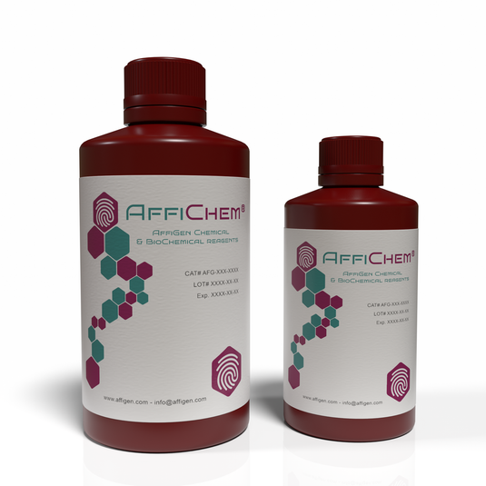
Introducing: Melzer Reagent (For Mycology)
A Foundational Staining Solution for Fungal Identification
After understanding its deep relevance in microscopy, taxonomy, fungal histology, ecological biology, and laboratory workflows, Melzer Reagent naturally remains one of the most essential solutions for mycologists.
Key Advantages
-
Reliable amyloid, dextrinoid, and inamyloid testing
-
Enhanced visualization of spores, asci, hyphae, cystidia
-
Supports taxonomy across Basidiomycota and Ascomycota
-
Compatible with educational, research, and environmental labs
-
Complements molecular and morphological identification
Conclusion
Melzer Reagent continues to be a foundational analytical tool in mycology. Its capacity to reveal iodine-reactive structures provides insight into fungal cell-wall chemistry and helps distinguish species with remarkable precision. Whether used for taxonomic research, biodiversity documentation, ecological evaluation, or student training, Melzer Reagent remains a gold standard for microscopic fungal identification.